Home
/ Abdominal Anatomy Diagram - Male Anatomy From The Back In 2021 Human Body Organs Anatomy Organs Human Anatomy Female : It is a flexible, dynamic container, housing most of the organs of the alimentary system and part of the urogenital system.
Abdominal Anatomy Diagram - Male Anatomy From The Back In 2021 Human Body Organs Anatomy Organs Human Anatomy Female : It is a flexible, dynamic container, housing most of the organs of the alimentary system and part of the urogenital system.
Abdominal Anatomy Diagram - Male Anatomy From The Back In 2021 Human Body Organs Anatomy Organs Human Anatomy Female : It is a flexible, dynamic container, housing most of the organs of the alimentary system and part of the urogenital system.. He has been with healthiack.com since 2012 and has written and reviewed well over 500 coherent articles. Anatomy and sonography of the abdominal vasculature diagram. It is a flexible, dynamic container, housing most of the organs of the alimentary system and part of the urogenital system. Is a health blogger focusing on health, beauty, lifestyle and fitness topics. Three unpaired branches of the abdominal aorta supply the organs of the digestive system.
Picture of abdomen the abdominal cavity is the part of the body that houses the stomach, liver, pancreas, kidneys, gallbladder, spleen, and the large and small intestines. In the rear of the abdomen are the back muscles and spine. At the level of the pelvic bones, the abdomen. This mri abdomen axial cross sectional anatomy tool is absolutely free to use. It can develop a bubble of sorts, which can be compared visually to a snake that swallowed a rat—sort of bloated in one spot.
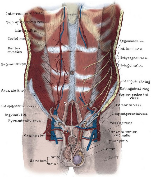
Anatomy Of Female Abdomen And Pelvis Trialexhibits Inc from cdn.trialexhibitsinc.com Lymphoid anonymous contributor abdominal muscles id 162 spare tire around their abdomen: The muscles of the abdomen protect vital organs underneath and provide structure for the spine. These muscles help the body bend at the waist. It is a flexible, dynamic container, housing most of the organs of the alimentary system and part of the urogenital system. The abdomen allows a lot of room for a weak aorta to expand and grow. This diagram depicts abdominal anatomy.human anatomy diagrams show internal organs, cells, systems, conditions, symptoms and sickness information and/or tips for healthy living. An intact woman's figure has space between the rib cage and the hip bones commonly known as the waist. Four abdominal quadrants and nine abdominal regions in anatomy and physiology, you'll learn how to divide the abdomen into nine different regions and four different quadrants.
We are pleased to provide you with the picture named the peritoneum anatomy diagram.
Learn vocabulary, terms, and more with flashcards, games, and other study tools. Is a health blogger focusing on health, beauty, lifestyle and fitness topics. For more anatomy content please follow us and visit our website: Vascular anatomy of the abdomen. • abdominal walls • abdominal cavity • abdominal viscera The abdomen allows a lot of room for a weak aorta to expand and grow. Three unpaired branches of the abdominal aorta supply the organs of the digestive system. Picture of abdomen the abdominal cavity is the part of the body that houses the stomach, liver, pancreas, kidneys, gallbladder, spleen, and the large and small intestines. Inflammation of the covering of the abdominal structures causing rigidity and severe pain. He has been with healthiack.com since 2012 and has written and reviewed well over 500 coherent articles. It is a flexible, dynamic container, housing most of the organs of the alimentary system and part of the urogenital system. Diagram the liver has more than 500 functions. Four abdominal quadrants and nine abdominal regions in anatomy and physiology, you'll learn how to divide the abdomen into nine different regions and four different quadrants.
For more anatomy content please follow us and visit our website: Four abdominal quadrants and nine abdominal regions in anatomy and physiology, you'll learn how to divide the abdomen into nine different regions and four different quadrants. The muscles of the abdomen protect vital organs underneath and provide structure for the spine. This all changes after hysterectomy. The muscles of the abdomen protect vital organs underneath and provide structure for the spine.
Anatomy Of The Lower Urinary Tract And Male Genitalia Abdominal Key from abdominalkey.com Lecture 4 abdominal vasculature gross anatomy unit 7. Spleen is the biggest lymphoid organ present in the upper far left portion of the abdomen in the left hypochondrium and is surrounded by peritoneum. The muscles of the abdomen protect vital organs underneath and provide structure for the spine. Using a 35 or 5 mhz curved array transducer place the transducer in a sagittal plane at the midline of the body just inferior to the xiphoid process of the sternum. This diagram depicts abdominal anatomy.human anatomy diagrams show internal organs, cells, systems, conditions, symptoms and sickness information and/or tips for healthy living. We hope this picture organs of abdomen diagram can help you study and research. 84 anatomical diagrams and histological images. We focused especially on the diagrams of the abdominal digestive system (oesophagus is described on the modules about the thorax and oral cavity/pharynx on the ent modules).
Thoracic duct (duck) is between 2 gooses, azy gous and esopha gus.
At the level of the pelvic bones, the abdomen. Anatomy and sonography of the abdominal vasculature diagram. The abdomen allows a lot of room for a weak aorta to expand and grow. Anatomynote.com found organs of abdomen diagram from plenty of anatomical pictures on the internet. The muscles of the abdomen protect vital organs underneath and provide structure for the spine. The abdominal quadrants can create a differential diagnosis for the cause, along with ot We are pleased to provide you with the picture named the peritoneum anatomy diagram. It is a flexible, dynamic container, housing most of the organs of the alimentary system and part of the urogenital system. In this image, you may find the peritoneum anatomy diagram. The abdomen colloquially called the belly tummy or midriff is the part of the body between the thorax chest and pelvis in humans and in other vertebrates. Picture of abdomen the abdominal cavity is the part of the body that houses the stomach, liver, pancreas, kidneys, gallbladder, spleen, and the large and small intestines. Use the mouse scroll wheel to move the images up and down alternatively use the tiny arrows (>>) on both side of the image to move the images.>>) on both side of the image to move the images. Signs and symptoms of an abdominal aortic aneurysm are back pain, deep abdominal discomfort, and possibly a pulsating mass in the abdomen.
Ligament and skeletal changes post hysterectomy. Thoracic duct (duck) is between 2 gooses, azy gous and esopha gus. This diagram depicts abdominal oblique muscle anatomy.human anatomy diagrams show internal organs, cells, systems, conditions, symptoms and sickness information and/or tips for healthy living. The medical information on this site is provided as an information resource only, and is not to be used or relied on. Three unpaired branches of the abdominal aorta supply the organs of the digestive system.
Abdomen Illustrations Visualisations Of Human Anatomy from www.medical-artist.com He has been with healthiack.com since 2012 and has written and reviewed well over 500 coherent articles. Relation to azygous vein and esophagus id 26 the duck between 2 gooses: An intact woman's figure has space between the rib cage and the hip bones commonly known as the waist. Use the mouse scroll wheel to move the images up and down alternatively use the tiny arrows (>>) on both side of the image to move the images.>>) on both side of the image to move the images. Four abdominal quadrants and nine abdominal regions in anatomy and physiology, you'll learn how to divide the abdomen into nine different regions and four different quadrants. The major muscles of the abdomen include the rectus. • abdominal walls • abdominal cavity • abdominal viscera At the level of the pelvic bones, the abdomen.
Inflammation of the covering of the abdominal structures causing rigidity and severe pain.
Transversus abdominis internal abdominal oblique rectus abdominis external abdominal oblique Learn vocabulary, terms, and more with flashcards, games, and other study tools. Using a 35 or 5 mhz curved array transducer place the transducer in a sagittal plane at the midline of the body just inferior to the xiphoid process of the sternum. The major muscles of the abdomen include the rectus. The enlargement of spleen is referred to as splenomegaly. The muscles of the abdomen protect vital organs underneath and provide structure for the spine. Abdomen and digestive system anatomy: Abdomen the abdomen is the part of the trunk between the thorax and the pelvis. • abdominal walls • abdominal cavity • abdominal viscera The diaphragm forms the upper surface of the abdomen. Ligament and skeletal changes post hysterectomy. Signs and symptoms of an abdominal aortic aneurysm are back pain, deep abdominal discomfort, and possibly a pulsating mass in the abdomen. We focused especially on the diagrams of the abdominal digestive system (oesophagus is described on the modules about the thorax and oral cavity/pharynx on the ent modules).
The enlargement of spleen is referred to as splenomegaly abdominal anatomy. For more anatomy content please follow us and visit our website:

